Muscle Imaging: Artificial Intelligence, Automatic Segmentation and Imaging Data Sharing in Neuromuscular Disease
- Number 286
- Date 7 March 2025
286th ENMC International Workshop
Location: Hoofddorp, The Netherlands
Title: Muscle Imaging: Artificial Intelligence, Automatic Segmentation and Imaging Data Sharing in Neuromuscular Disease
Date: 7-9 March 2025
Organisers: Prof. V. Straub (UK), Dr H. Kan (Netherlands), Dr J. Warman Chardon (Canada), Prof. J. Vissing (Denmark).
Early Career Researchers (ECR): Dr Sarah Schläger (Germany), Dr Susi Rauh (The Netherlands)
Translations of this report by:
German by Dr Sarah Schläger
French by Dr Pierre Carlier
Italian by Dr Mauro Monforte
Danish by Prof. John Vissing
Dutch by Dr Susi Rauh
Russian by S. Mitlenok
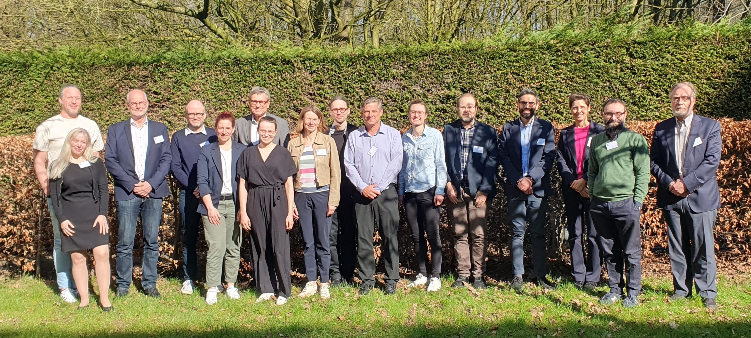
Participants: Prof. Volker Straub (United Kingdom), Dr Hermien Kan (The Netherlands), Dr Jodi Warman-Chardon (Canada), Prof.John Vissing (Denmark), Dr Francesco Santini, (Switzerland), Dr Harmen Reyngoudt (France), Dr Lara Schlaffke (Germany), Dr Mauro Monforte (Italy), Dr Glenn Walter (United States), Prof. Giorgio Tasca (United Kingdom), Prof. Anna Pichiecchio (Italy), Dr Martijn Froeling (The Netherlands), Dr Jasper Morrow (United Kingdom), Prof. John Thornton (United Kingdom), Dr Pierre Carlier (Belgium), Prof. Kristl Claeys (Belgium), Dr Sarah Schläger (Germany), Dr Susi Rauh (The Netherlands).
Background Information
In neuromuscular diseases (NMDs), muscles can lose volume, can be replaced by fat or be affected by inflammation. In many NMDs, these changes affect different muscles to different degrees, leading to selective and sometimes very specific patterns of muscle abnormalities. Quantitative muscle magnetic resonance imaging (qMRI) can measure muscle volume, fatty, inflammatory, necrotic or dystrophic changes objectively and non-invasively. This makes qMRI a useful imaging biomarker for diagnosing NMDs, tracking disease progression, and evaluating treatment response.
To use qMRI effectively in everyday medical practice, clinicians and researchers need consensus to identify the specific qMRI techniques to perform, accurate methods for identifying each muscle, and standardized ways to report the results. One of the biggest challenges is identifying each muscle in the MR images, known as “muscle segmentation”. Muscle segmentation is essential to identify muscle patterns of involvement and progression of disease. Manual muscle segmentation is too time-consuming for routine clinical use and recently, computer-based tools using artificial intelligence (AI) have now been developed to automate this process. Nevertheless, there is no agreement yet on the best method or tool to use. Additionally, large collections of MRI scans are needed from multiple medical centers for training and testing to improve these AI tools.
The goal of this workshop was to agree on standard methods for performing qMRI scans, analyzing muscle images, and reporting findings. This will help bringing qMRI from research settings into everyday clinical practice to improve patient care.
Workshop Aims
- qMRI Image Acquisition: The goal is to agree on standard practices for performing qMRI scans and storing the images, through discussions involving radiologists, physicists, and neurologists.
- qMRI Data Analysis: The aim is to set standards for processing, analyzing, and describing the data from these qMRI studies. This includes deciding how to store the data and how to incorporate new machine learning techniques into the analysis.
- Automatic Segmentation: The focus is to agree on standards for processing, analyzing, and describing the data from automatic muscle segmentation in qMRI studies.
- Data Sharing: The objective is to build stronger collaboration between centers in Europe and around the world, to improve strategies for sharing imaging data and ensuring its long-term sustainability.
Workshop Outcomes and Deliverables
The experts agreed on a standard procedure for performing qMRI scans to diagnose NMDs, specifically to measure fat content as an imaging biomarker and water-T2 values as an imaging biomarker of disease activity. The data from these scans will be stored in a standardized format (ORMIR-MIDS), making it easier to compare results across different locations and studies and simplifying the analysis and sharing of qMRI data. For diagnostic purposes, segmenting individual muscles is necessary, whereas grouping muscles together may be useful for tracking disease progression.
The experts also recommend investing in machine-learning resources to implement automatic muscle segmentation in clinical practice. More research is needed to compare different automatic segmentation methods and to ensure accuracy of the segmentations for various NMDs. Therefore, organising an international challenge to compare muscle segmentation techniques will be pursued. To aid the implementation of qMRI-based biomarkers in clinics, a standardized format for radiological reporting will be created.
Impact for the patients and their families
Improving standards for performing and analyzing qMRI scans will help clinicians and researchers diagnose specific NMDs and assess disease progression more accurately. This will also result in better treatment monitoring, help in understanding disease mechanisms, and will enable comparison of results from different studies across the world.
Next steps
- An automatic muscle segmentation challenge will be held to test different methods and tools for automatically identifying muscles in qMRI scans.
- A structured radiological report form will be designed to help integrate qMRI in routine clinical practice.
A full report will be published in Neuromuscular Disorders (PDF).

