Muscle Imaging in Facioscapulohumeral Muscular Dystrophy (FSHD): relevance for clinical trials
- Number 265
- Date 22 April 2022
Location: Hoofddorp, The Netherlands
Title: 265th ENMC International Workshop “Muscle Imaging in Facioscapulohumeral
Muscular Dystrophy (FSHD): relevance for clinical trials”
Date: 22 – 24 April 2022
Organizers: Giorgio Tasca (Italy), Shahram Attarian (France), John Vissing (Denmark), Jordi Diaz-Manera (UK).
Translations of this lay report:
French by Prof. Robert Carlier
Dutch by Dr Sanne Vincenten
German by Dr Teresa Gerhalter
Italian by Mauro Monforte
Danish by Prof. John Vissing
Polish by Michal Rataj
Spanish by María Gómez-Rodulfo
Participants: Hermien Kan (the Netherlands), Nens van Alfen (the Netherlands), Anna Pichiecchio (Italy), Pierre Carlier (France), Robert-Yves Carlier (France), Sabrina Sacconi (France), Roberto Fernandez Torron (Spain), Francesco Santini (Switzerland), Teresa Gerhalter (Germany), David Bendahan (France), Doris Leung (USA), Linda Heskamp (UK), Kristen Meiburger (Italy), Nicol Voermans (the Netherlands), Aurea Martins-Bach (UK), Olof Dahlqvist Leinhard (Sweden).
Mauro Monforte (Italy) and Sanne Vincenten (the Netherlands) in the Early-Career Programme. Maria Vriens-Munoz Bravo (the Netherlands), Raj Badiani (UK) and Michal Rataj (Poland) as patient representatives. George Padberg (the Netherlands) attended as listener.
The 265th ENMC International Workshop on “Muscle Imaging in Facioscapulohumeral
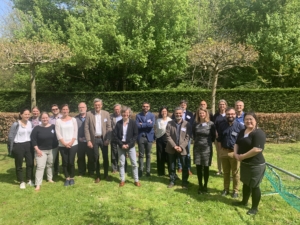
Muscular Dystrophy (FSHD): relevance for clinical trials” took place as a hybrid meeting, with 21 participants on site and 5 connected remotely.
Facioscapulohumeral muscular dystrophy (FSHD), one of the most frequent muscular dystrophies, is at the doorstep of clinical therapeutic trials. The scientific community is committed to reach clinical trial readiness, and two major consortia devoted to boost drug development have been created (the FSHD Clinical Trial Research Network, based in the US, CTRN, and more recently the FSHD European Trial Network, ETN). Notably, FSHD is unique in its genetic mechanism and very peculiar in the progression of muscle damage compared to the other muscular dystrophies.
Muscle imaging through magnetic resonance (MRI) has lately been established as an important tool to diagnose and follow the evolution of different neuromuscular disorders. In FSHD, evidence derived from MRI studies substantially contributed to a better understanding of this disease and its variable progression over time. However, a major need not yet addressed is the clear establishment of the importance of muscle imaging for the diagnosis and follow-up of FSHD patients, and the definition of its role in a clinical trial setting. This workshop provided a unique opportunity to gather the experts in the field, who shared their knowledge and experiences, and an unprecedented occasion to have dedicated time to focus and discuss on the usefulness and harmonization of the imaging techniques specifically in FSHD, since no previous meeting has been specifically devoted to address these issues so far.
As a preparatory activity of the workshop a survey of the imaging facilities (MRI, ultrasound) available at each participating center and used for evaluation of FSHD had been disseminated and the results will be part of the deliverables.
Day 1. Session 1 on the use of “Qualitative MRI” was chaired by Giorgio Tasca and focused on the application of conventional/standard MRI sequences (mainly T1w and T2w-STIR) to address diagnostic challenges and to give clues about disease activity and progression, which is particularly heterogeneous in this disease. The experience of the different neuromuscular centers in Italy, France, and Spain was presented. The possibility to derive similar information from the visual evaluation of quantitative MRI protocols (Dixon), the differences found in FSHD type 1 vs type 2 patients, as well as gender differences were discussed.
Day 2. Session 2 (“Quantitative MRI”, chaired by John Vissing) dealt with the application of MRI protocols capable of measuring the fat content and the water dynamics of the individual muscles. The experience of different centers (located in Denmark, Italy, the Netherlands and USA) with the use of such techniques in cross-sectional and longitudinal cohorts of FSHD patients was presented. Common findings and differences in acquisition protocols were discussed, and a proposal of a shared analysis of data was made as an action point of the workshop. The possibility to have whole-body fat and water quantitative information in short timeframes using different protocols, whose application can be tailored depending on the tradeoffs between multicenter implementation and technical resources available at the different centers, was also presented.
Session 3 (“Experience from trials using MRI”) was chaired by Shahram Attarian. Muscle MRI has already been used in the context of two international multicenter interventional trials on FSHD (ATYR1940 from aTyr pharma and Redux4 from Fulcrum therapeutics). The involved participants shared their experience and highlighted how MRI biomarkers were implemented, both for enrolment and as outcomes, and what lessons were learned, also compared with similar experiences in other neuromuscular disorders. Olov Dahlqvist Leinhard (representative of the company AMRA medical, connected remotely for the time of the talk and the following discussion) illustrated their protocol, the analysis pipeline and the different MRI-derived biomarkers regarding muscle volume and fat content analysis that were used in the ReDux4 trial.
Session 4 on “Correlation with functional outcomes and other techniques” was chaired by Jordi Diaz-Manera. The clinical relevance of the imaging measurements was addressed by a detailed analysis of the evidence available. Data suggested that quantitative MRI is able to detect changes in the muscle structure before patients experience clinical decline, and that some metrics might be helpful to predict changes in muscle function over time. Artificial intelligence powered methods for muscle segmentation, which is an essential but currently time-consuming step needed for quantitative imaging, were also presented. Innovative techniques able to detect fibrosis and MRI spectroscopy were the topics of the last presentations of this session.
Finally, Session 5 on “Muscle Ultrasound” was chaired by Nens van Alfen. Ultrasound is a non-invasive technique able to visualize muscles and assess their structural features. A detailed protocol used for the study of neuromuscular patients, and FSHD in particular, was presented and discussed among participants, with a general interest in possible application at different centers. Cross-sectional and longitudinal ultrasound data, also for the facial muscles, which are difficult to assess by other imaging techniques, were shown by the Dutch group. Deep learning for automatic ultrasound muscle segmentation, in parallel with what was presented in the previous session, was the topic that closed the works of the day.
Day 3 was fully dedicated to the general discussion. The participants agreed on the diagnostic usefulness of MRI in particular contexts, and on its role in patients’ stratification to enter a clinical trial. Ultrasound expertise is currently restricted to specific centers, but emerges as an interesting technique that, if standardized in multiple sites, could help in providing additional information to MRI also in a clinical trial framework.
Concerning quantitative MRI, efforts should be made to harmonize and improve protocols that had been already implemented in previous trials. Researchers should aim to achieve a whole-body coverage given the heterogeneity and unpredictability of FSHD, as well as to include specific sequences to assess disease activity. The idea to tailor the choice of the specific imaging biomarker(s), based on the action of the investigated drug, also clearly emerged.
A consensus was reached towards the need to perform a global analysis of the quantitative MRI data published in the different natural history studies, with the aims to increase validity and possibly obtain additional insights on the disease mechanism and progression. Steps towards the implementation of the new advanced imaging protocols, which could provide more complete information in a shorter acquisition time, should be pursued. A coordination with the imaging working group already in place in the CTRN is mandatory to optimize efforts, and joint meetings, both online and live, will be arranged for this purpose.
To disseminate the results of the workshop, they will be presented at the next FSHD ETN meetings and at the 2022 FSHD IRC congress, in addition to the full report that will be published in Neuromuscular Disorders.
The full report is published in Neuromuscular Disorders (PDF)

